Table of Contents
Webinar Video
Please note, this is the second webinar David has done for CAUK, but this is the third blog post. We apologise for any confusion.
Introduction
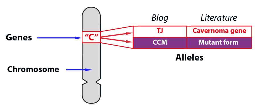
In the first part of this series, explained that cavernoma leak blood because the tight junctions between blood capillary cells are lost. In this blog we will explain what happens inside the cells to allow this.
3A. Tight Junction Recap
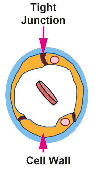
In the first part of the series, we showed that a crititcal difference between normal blood capillaries in the brain and the cavernoma is the presence or absence of Tight Junctions that hold the cells tightly together so that material cannot pass across the capillary cell wall.
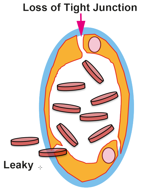
3C. CCM Genes code for the Cavernoma Adaptor Complex
When the three known ‘cavernoma’ genes all have the ‘Tight Junction [TJ]’ form, the resulting three proteins form a complex
known as the Adaptor CCM Complex and no cavernoma can form.
Complex
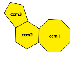
The presence of a CCM form of one of the genes results in no Adaptor CCM Complex format and a cavernoma can form.
No Complex

3D. Tight and Leaky Junctions
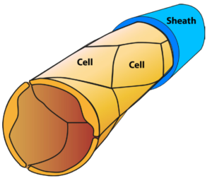
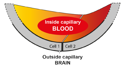
Figure 1 shows a capillary cell with a cut end, and Figure 2 an enlarged image of that cut end with a junction between two cells.
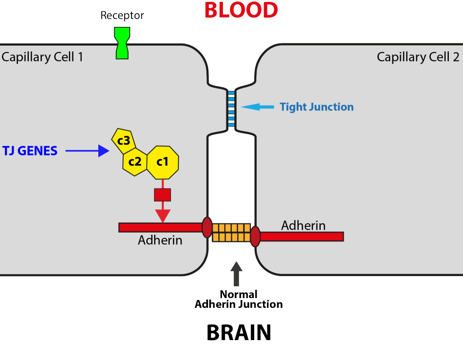
Figures 3 and 4 are further enlargements to show the junction between the cells in more detail, including just those components of the cell needed to explain the maintenance and loss of tight junctions.
Tight Junction genes: no cavernoma (Figure 3)
This is the normal situation found in over the 99.9% of the population without a cavernoma. The tight junction is maintained by the protein adherin. This spans the cell membrane with a part inside the cell (red) and a part outside (orange).
The conformation (shape and properties) of adherin are controlled by the Adaptor CCM Complex protein (yellow) via other components shown by the red arrow.
The external part of adherin binds tightly to the external component of the adjacent cell. A second element of the tight junction is a set of other tight junction proteins
shown in blue.
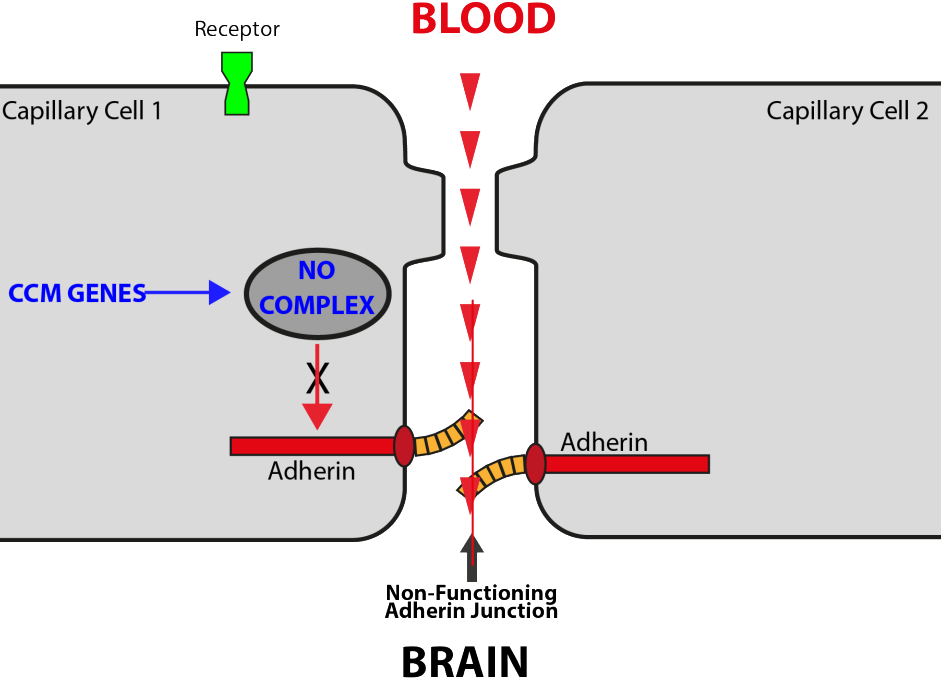
CCM genes: cavernoma possible (Figure 4)
As described in Blog 2, when both copies of any one of your three cavernoma genes in a capillary cell have the CCM form, the Adaptor CCM Complex is lost and so cannot exert its control.
The link to the adherin molecule is absent and the adherin molecule loses its ability to bind to the adherin from the adjacent cell.
The tight junction proteins (blue) no longer span the junction and blood can pass from the blood capillary directly to the brain.
The capillary thus become leaky and a cavernoma can begin to form.
![]() This content is provided under the Creative Commons Attribution-NonCommerical-NoDerivatives License.
This content is provided under the Creative Commons Attribution-NonCommerical-NoDerivatives License.
The full license is available here, and a human readable version is available here.


